
Be a NURSE Your Country Needs You
Cart (0)

Breech Presentation: Definition,Type, Etiology and Mechanism for 3rd Year GNM A Complete Note

Definition of Breech Presentation:

Breech presentation is a fetal position during childbirth where the baby’s buttocks, feet, or both are positioned to emerge first, instead of the head.

In breech presentation, the lie is longitudinal and the podalic pole presents at the pelvic brim. It is the
most common malpresentation.

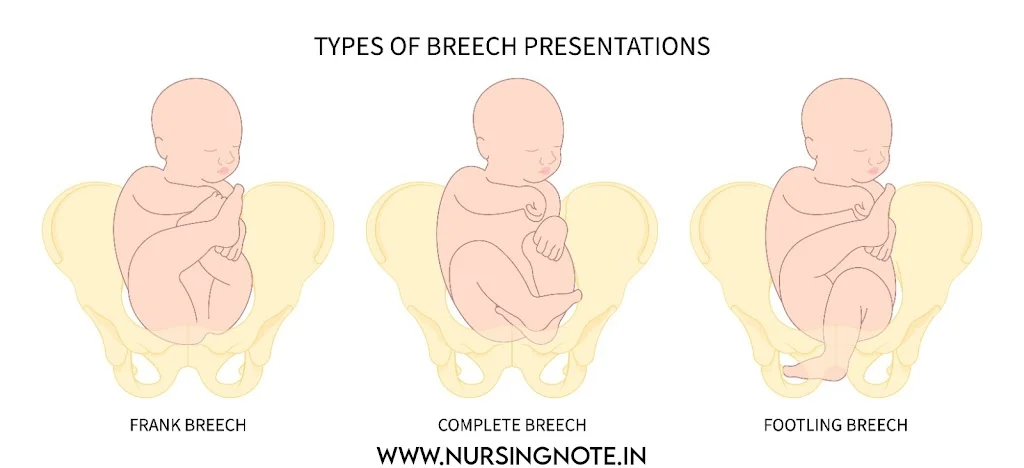
Frank Breech, Illustration of breech fetal positions: complete, incomplete, and footling breech.Complete Breech, Footling Breech, Kneeling Breech
Breech Presentations

Types of Breech Presentation

There are two varieties of breech presentation:
 

  • Complete
  • Incomplete

1. Complete Breech (Flexed Breech):

In a complete breech, the fetus maintains the normal attitude of full flexion.

  • Position: Thighs are flexed at the hips, and legs are flexed at the knees.
  • Presenting Part: Two buttocks, external genitalia, and both feet present at the birth canal.
  • Prevalence: This type is more common in multiparous women (those who have had previous deliveries), with an occurrence of about 10%.

2. Incomplete Breech:

An incomplete breech results from varying degrees of extension of the thighs or legs at the fetal pole. This type includes three distinct varieties:

A.Frank Breech:

  • Position: Thighs are flexed on the trunk, while legs are extended at the knee joints.
  • Presenting Part: Two buttocks and external genitalia only (without the feet).
  • Prevalence: The most common form of breech, especially in first-time pregnancies (primigravidae), accounting for around 70%. This is attributed to the firmer abdominal wall and higher uterine tone in primigravida, facilitating early engagement of the breech.

B. Footling Presentation:

  • Position: Both thighs and legs are partially extended, causing one or both feet to present at the birth canal.
  • Prevalence: Occurs in about 25% of breech presentations. Footling breech poses a higher risk for complications such as cord prolapse.

C. Knee Presentation:

  • Position: Thighs are extended, but knees are flexed, causing the knees to present at the birth canal.
  • Prevalence: This is the least common form of breech presentation

Etiology of Breech Presentation

  1. Prematurity: The most common cause, as the fetus may not have had time to turn head-down.
  2. Breech with Extended Legs: Extended legs make it difficult for the fetus to rotate.
  3. Twins: Limited space in multiple pregnancies restricts fetal movement.
  4. Oligohydramnios: Low amniotic fluid reduces fetal mobility.
  5. Congenital Uterine Abnormalities: Abnormalities such as a septate or bicornuate uterus restrict fetal movement.
  6. Short Umbilical Cord: A short cord limits fetal movement.
  7. Hydrocephalus: A large fetal head can fit better in the wide fundal area of the uterus.
  8. Placenta Previa: A low-lying placenta blocks the fetus from turning head-down.
  9. Contracted Pelvis: A smaller pelvic opening restricts fetal rotation.
  10. Hydramnios: Excessive amniotic fluid allows more fetal movement.
  11. Multiparity: Lax abdominal walls in women who have had multiple pregnancies increase fetal mobility.
  12. Genetic Conditions: Trisomies (13, 18, 21) can affect fetal muscle tone.

Diagnosis of Breech Presentation

A. Clinical Diagnosis:

Abdominal Palpation: Through Leopold’s maneuvers, the healthcare provider may detect the fetal head at the upper part of the uterus (fundus) and the buttocks or feet near the pelvic inlet.
Vaginal Examination: If labor has begun, a vaginal exam may reveal the fetal buttocks, feet, or genitalia as the presenting part instead of the head.
Auscultation: The fetal heartbeat is often heard above the mother’s umbilicus in breech presentation, rather than lower in the pelvis, as is typical in head-down (cephalic) presentations.

B. Sonography (Ultrasound):

Confirms the fetal position, assesses factors like amniotic fluid, placental location, and potential fetal abnormalities related to breech presentation.

Mechanism of Labor in Breech Presentation

In breech delivery, the primary movements occur in sequence for the buttocks, shoulders, and head:

A. Buttocks:

  • Engagement: The buttocks engage in an oblique diameter of the pelvic inlet, with the bitrochanteric diameter (10 cm) leading.
  • Descent: The buttocks descend until the anterior buttock reaches the pelvic floor.
  • Internal Rotation: The anterior buttock rotates behind the symphysis pubis.
  • Further Descent and Lateral Flexion: The anterior hip passes under the symphysis pubis, followed by the posterior hip, allowing trunk and lower limbs to deliver.
  • Restitution: The buttocks rotate back to their original oblique position.

B. Shoulders:

  • Engagement: The bisacromial diameter (12 cm) engages in the same oblique diameter as the buttocks.
  • Descent and Internal Rotation: Shoulders rotate into the anteroposterior diameter of the pelvic outlet as the trunk externally rotates.
  • Delivery: The posterior shoulder emerges first, followed by the anterior shoulder through flexion of the trunk.
  • Restitution and External Rotation: The shoulders rotate, aligning the fetal trunk in a dorsoanterior position.

C. Head:

  • Engagement: The head engages either in the opposite oblique or transverse diameter, with the suboccipitofrontal diameter (10 cm) leading.
  • Descent with Flexion: The head descends with increasing flexion.
  • Internal Rotation: The occiput rotates anteriorly, positioning behind the symphysis pubis.
  • Descent and Flexion for Delivery: The subocciput hinges under the symphysis pubis, and the head delivers by flexion, with the chin, mouth, nose, forehead, and occiput appearing successively.
  • Expulsion: Expulsion of the head relies on maternal bearing-down efforts, not on uterine contractions.

FAQs Section

  • The Most Frequently Asked Question related Breech Presentation are:
    • Q1: What is the most common type of breech presentation?
      A1: The frank breech is the most common, occurring in about 70% of cases.
    • Q2: What are the risks of breech delivery?
      A2: Risks include cord prolapse, head entrapment, and delivery complications.

Fetal Circulation – Definition, Umbilical Cord and Fetal Circulation Flow Chart

Diagram of fetal circulatory system

 FETAL CIRCULATION

➦ Definition:

Fetal Circulation or Foetal Circulation refers to the unique circulatory system that allows the exchange of gases and nutrients between the developing fetus and the pregnant mother.


A picture of fetal circulation
Fetal Circulation Diagram


➦ Umbilical Cord:

        ● Umbilical Arteries:

Return non-oxygenated blood, Biological waste, and CO₂ from fetal body to placenta.

        ● Umbilical Vein:

Bring Oxygenated blood and nutrients to the fetus.

The umbilical vein carries 80% saturated blood from the placenta.

➦ Shunts:

       ● Ductus Venosus:

Connects the umbilical vein and inferior vena cava.

       ● Ductus Arteriosus:

Connects the main pulmonary artery to the aorta.

       ● Foramen Ovale:

Anatomical opening between right and left atrium.

➦ Cardiac Output:

  • During fetal life – 350 ml/kg/min
  • Following birth – 500 ml/kg/min

➦ Fetal Heart Rate;

The normal fetal heart rate is 120 – 160 beats/min.

➦ Fetal Circulation Flow Chart:

 
A picture of fetal circulation steps
Fetal Circulation Flow Chart
 

➦ Fetal Circulation Explained Video:


➦ Download NOTE of Fetal Circulation:

What is Uterine Fibroids? – Types, Clinical Features, Symptoms, Diagnosis and Treatment

Educational overview of uterine fibroids.

Uterine Fibroids

Definition of Fibroid:

Fibroid tumors are benign tumors arising in the myometrium which can protrude into the uterine cavity, bulge through the outer layer or grow within the myometrium.

Uterine Fibroids - Definition, Types,Signs and Symptoms, Diagnosis and Treatment

Growth of Fibroids

1. Fibroids develop due to proliferation of smooth muscle cells. The tumors usually grow very slowly, about 1 to 2 cm per year.

2. Fibroids are estrogen dependent and thus grow rapidly during pregnancy. They do not occur before menarche and growth cessation occurs after menopause.

Types of Fibroids:

Alternatively named myomata or leiomyomata, uterine fibroids are the commonest of all pelvic tumors. They are benign fibromuscular swellings, arising in the muscle wall of the uterus. Fibroids are oestrogen sensitive.

➦ Submucous: It lies immediately below the endometrium and enlarges the surface of the uterine cavity often leading to menorrhagia. Fibroids may become pedunculated forming polypi which can extrude through the cervix.

➦ Intramural: The commonest site for fibroids, surrounded by smooth muscle, enlarging the uterine wall and distorting venous drainage.

➦ Subserous: Fibroids just beneath the peritoneum on the outer uterine surface. May be on an elongated stalk (pedunculated) with a risk of torsion; may grow into the broad ligament.

Clinical Features of Fibroid:

1. Majority of fibroids (75%) remains asymptomatic and are detected during routine examination or at laparotomy or laparoscopy.

2. The uterus reaches a 4 to 5 months gestation size without symptoms in most cases.

3. In symptomatic type, menorrhagia is the classic symptom.

4. Metrorrhagia or irregular bleeding is seen in some women.

5. Dysmenorrhea is a symptom when it is associated with pelvic congestion or endometriosis.

6. Infertility is seen in about 30 percent of women.

7. Recurrent pregnancy loss (Miscarriage or preterm labor).

8. Lower abdominal or pelvic pain.

Symptoms of Fibroid:

    • None: a pelvic swelling is found incidentally on examination.
    • Occasional tightness of waistband of clothes.
    • Pressure: bladder compression causing daytime frequency and occasionally impaired urinary stream. In the supporting ligaments it causes backache and overall sensation of pelvic heaviness.
    • Pain: associated with red degeneration or torsion of subserous pedicles. Dysmenorrhoea may indicate the presence of a submucous fibroid.
    • Menstrual disturbances:

a. Menorrhagia: heavy bleeding;
b. Metrorrhagia: prolonged menses;
c. Irregular, intermittent bleeding: often associated with polyps and other surface lesions.

Diagnosis of Fibroid:

1. Majority of uterine fibroids can be diagnosed from S history and pelvic examination

2. Ultrasound and color Doppler are used to locate the fibroids accurately.

3. Laparoscopy is useful if the uterine size is less than 12 weeks and associated with pelvic pain and infertility

4. Uterine curettage is done in the presence of irregular bleeding to detect any coexisting pathology and to study the endometrial pattern.

Differential diagnosis

  • Pregnancy: particularly if fibroids have been softened by cystic degeneration.
  • Ovarian tumor: often cystic, unilateral and does not move with cervical displacement.
  • Adenomyosis: more commonly causes uniform diffuse and tender uterine enlargement.

Management of Fibroid:

Therapeutic intervention depends on the woman’s desire for future pregnancy and the tumor size.

Medical management:

Hormone therapy may be used as a short-term intervention to decrease the size of fibroid and to minimize blood loss. Drugs used are antifibrinolytics, antiprogesterones, Danazol, GnRH analogous and prostaglandin synthetic inhibitors. Use of hormones for more than 6 months can increase the risk of osteoporosis.

●    If small and asymptomatic, conservative management with annual examination and ultrasound monitoring of size is sufficient. This is especially used in women over 40 because fibroids do not grow after menopause and may shrink.

●    Menstrual or pressure symptoms may dictate surgery.

●    Pain-requires analgesia.

●    Heavier and longer periods with anemia are the commonest indication for proceeding to surgery.

●    Embolization under radiological control: A cannula is passed into the uterine arteries via the femoral artery. The uterine arteries are embolized by injecting tiny silicon particles causing the fibroids to degenerate. Pain relief is essential for 48 hours. In women with large/multiple fibroids there is an increased risk of hysterectomy compared to surgical treatment. Pyrexia and abscess forination can occur. It is not recommended for women who wish to preserve their fertility, although live births have been reported following the procedure.

Surgical management :

    1. Abdominal hysterectomy:

Suitable when family is complete with women over the age of 40 or when the uterus is grossly enlarged and distorted by multiple fibroids.

    1. Vaginal hysterectomy:

When fibroids are small and few in number and there is an associated prolapse of the uterus.

    1. Myomectomy:

In young women whose families are incomplete or when there is a personal desire to retain the uterus. The procedure is often vascular and may cause scarring, with adhesion formation impairing fertility. If the fibroids are numerous it may be impossible to remove them all, and growth of the remainder may cause problems in the future. Women undergoing this procedure should be warned that the surgeon may have to proceed to a hysterectomy and consent to the possibility of this (1% risk).

Submucous fibroids may be resected with laser or diathermy through a hysteroscope.

Destructive Operations – Craniotomy, Evisceration, Decapitation and Cleidotomy

Destructive Operations

Definition:

The destructive operations are designed to diminish the bulk of the fetus so as to facilitate easy delivery through the birth canal. In modern obstetric practice, virtually there is hardly any place for destructive operations. Neglected obstetrics requiring destructive operations are completely preventable. These procedures are difficult and may be dangerous too unless the operator is sufficiently skilled. Unfortunately, one may have to perform such operations while working in the unorganized sector. Some commonly performed operations are discussed here. There are four types of operations:

♦Craniotomy             ♦Evisceration             ♦Decapitation             ♦Cleidotomy

CRANIOTOMY

DEFINITION: It is an operation to make a perforation on the fetal head, to evacuate the contents followed by extraction of the fetus.

INDICATIONS: 

■ Cephalic presentation producing obstructed labor with dead fetus: This is the most common indication  of craniotomy in the referral hospitals of the developing countries.

■ Hydrocephalus even in a living fetus: This is applicable both for the forecoming and the aftercoming head .

■ Interlocking head of twins.

CONDITIONS TO BE FULFILLED: (1) The cervix must be fully dilated and (2) baby must be dead (hydrocephalus being excluded).

CONTRAINDICATION: (i) The operation should not be done when the pelvis is severely contracted so as to shorten the true conjugate to less than 7.5 cm (3″). In such condition, the baby cannot be delivered, as the bimastoid diameter (base of the skull) of 7.5 cm cannot be compressed. (ii) Rupture of the uterus where laparotomy is essential.

PROCEDURES: Preliminaries: The preliminary preparations are the same as mentioned in p. 642. The operation is to be done under general anesthesia.

Actual steps

Step I: The two fingers (index and middle) are introduced into the vagina and the finger tips are to be placed on proposed site of perforation. However, when the suture line cannot be defined because of big caput, the perforation should be done through the dependent part.

Sites of perforation: Vertex: On the parietal bone either side of the sagittal suture. Suture is avoided to prevent collapse of the bone thereby preventing escape of the brain matter. Face: Through the orbit or hard palate. Brow: Through the frontal bone.

Step II: The Oldham’s perforator  with the blades closed is introduced under the palmar aspect of the fingers protecting the anterior vaginal wall and the adjacent bladder  until the tip reaches the proposed site of perforation.

Step III: By rotating movements the skull is perforated. During this step, an assistant is asked to steady the head per abdomen in a manner of first pelvic grip. After the skull is perforated, the instrument is thrust up to the shoulders and the handles are approximated so as to allow separation of the sharp blades for about 2.5 cm. The blades are again apposed by separating the handles. The instrument is brought out keeping the tip of the blades still inside the cranium. The instrument is rotated at right angle and then again thrust in up to the shoulders. The handles are once more to be compressed so as to separate the blades for about 2.5 cm. The perforated area now looks like a cross. The instrument with the blades closed is then thrust in beyond the guard to churn the brain matter. The instrument, with the blades closed, is brought out under the guidance of the two fingers still placed inside the vagina.

 Alternative to Oldham’s perforator, similar procedure could be performed using a sharp-pointed Mayo’s scissors. 

Step IV: With the fingers brain matter is evacuated. The idea is to make the skull collapse as much as possible.

Step V: When the skull is found sufficiently compressed, the extraction of the fetus is achieved either by using a cranioclast or by two giant volsella. Giant volsella are used to hold the incised skull and scalp margins.

PROCEDURES OF CRANIOTOMY

Step VI: The traction is now exerted in the same direction as like that mentioned in forceps operation. Step VII: After the delivery of the placenta, the uterovaginal canal must be explored as a routine for evidence of rupture uterus or any tear. Injection methergine 0.2 mg is to be given intravenously with the delivery of the anterior shoulder. The rest of the delivery is completed as in normal delivery.

Forceps versus craniotomy in a dead fetus: If the delivery of the uncompressed head can be accomplished without much force with consequent injuries to the mother, forceps delivery is preferred. But if it is found difficult and damaging to the mother, craniotomy is safer.

DECAPITATION

DEFINITION: It is a destructive operation whereby the fetal head is severed from the trunk and the delivery is completed with the extraction of the trunk and that of the decapitated head per vaginum. INDICATION:

(1) Neglected shoulder presentation with dead fetus where neck is easily accessible.

(2) Interlocking head of twins.

PROCEDURES: Preliminaries — The preliminaries to be followed are the same as outlined earlier. The operation is done under general anesthesia.

Actual Steps

Step I: If the fetal hand is not prolapsed, bring down a hand. A roller gauze is tied on the fetal wrist and an assistant is asked to give traction towards the side away from the fetal head to make the neck more accessible and fixed.

Step II: Two fingers of the left hand (middle and index) are introduced with the palmar surface downwards and the finger tips are to be placed on the superior surface of the neck—the proposed site of decapitation.

Step III: The decapitation hook with knife is to be introduced flushed under the guidance of the fingers placed into the vagina, the knob pointing towards the fetal head. The hook is pushed above the neck and rotated to 90° so as to place the knife firmly against the neck. The internal fingers, in the meantime, are placed on the under surface of the neck to guard the tip of the hook.

Step IV: By upward and downward movements of the hook with knife, the vertebral column is severed (evident by sudden loss of resistance). The rest of the soft tissue left behind may be severed by the same instrument or by embryotomy scissors. While removing the decapitation hook—it is to be pushed up; rotated to 90° and then to take out under the guidance of the internal fingers. The decapitated head is pushed up and the trunk is delivered by traction on the prolapsed arm.

Step V: Delivery of the decapitated head—Any of the following methods may be usually effective:

• By hooking the index finger into the mouth  • By holding the severed neck with giant vulsellum and delivery of the head as that of aftercoming head in breech  • Using forceps.

Step VI: Routine exploration of the uterovaginal canal to exclude rupture of the uterus or any other injury.

EVISCERATION

The operation consists of removal of thoracic and abdominal contents piecemeal through an opening on the thoracic or abdominal cavity at the most accessible site. The object is to diminish the bulk of the fetus which facilitates its extraction. If difficulty arises, the spine may have to be divided (spondylectomy) with embryotomy scissors.

The indications are: (1) Neglected shoulder presentation with dead fetus; the neck is not easily accessible and (2) fetal malformations, such as fetal ascites or hugely distended bladder or monsters.

CLEIDOTOMY

The operation consists of reduction in the bulk of the shoulder girdle by division of one or both the clavicles.The operation is done only in dead fetus (anencephaly excluded) with shoulder dystocia. The clavicles are divided by the embryotomy scissors or long straight scissors introduced under the guidance of left two fingers placed inside the vagina.

POSTOPERATIVE CARE FOLLOWING DESTRUCTIVE OPERATIONS

♦ Exploration of the uterovaginal canal must be done to exclude rupture of the uterus or lacerations on the vagina or any genital injury.

♦A selfretaining (Foley’s) catheter is put inside, especially following craniotomy for a period of 3–5 days or until the bladder tone is regained.

♦Dextrose saline drip is to be continued till dehydration is corrected. Blood transfusion may be given, if required.

♦Ceftriaxone 1 g IV infusion is given twice daily.

COMPLICATIONS: (1) Injury to the uterovaginal canal (2) rupture of uterus (3) postpartum hemorrhage—atonic or traumatic (4) shock—due to blood loss and/or dehydration (5) puerperal sepsis (6) subinvolution (7) injury to the adjacent viscera— bladder—vesicovaginal fistula or rarely to rectal wall leading to rectovaginal fistula and (8) prolonged ill health.

General Nursing and Midwifery (GNM) 3rd Year Suggestion – 2023

3rd Year GNM Nursing suggestions 2023

Suggestion for 3rd Year GNM Final Examination – 2023


Download the best PDF suggestion for GNM (General Nursing and Midwifery) 3rd year examination by Nursing Note with guaranteed common in the final examination.
 
General Nursing and Midwifery (GNM) 3rd Year Suggestion - 2023
 

📌 GNM Course details:

Aspect Details
Course Name General Nursing and Midwifery (GNM)
Duration 3.5 years (including internship)
Eligibility 10+2 with Science (Physics, Chemistry, Biology) or Arts with a minimum of 45% aggregate marks.
Admission Process Entrance exams or direct admission, depending on the institution
Curriculum – Anatomy and Physiology
– Fundamentals of Nursing
– Medical-Surgical Nursing
– Obstetric Nursing
– Pediatric Nursing
– Community Health Nursing
– Psychiatric Nursing, etc.
Internship Compulsory internship in hospitals and community health settings
Certification GNM certification upon successful completion
Career Opportunities Staff Nurse, Community Health Worker, Midwife, Nursing Educator, Nurse Administrator, etc.
Registration After completing the course, graduates need to register with the respective nursing council for practice.
Further Studies Opportunities to pursue B.Sc. Nursing or other advanced nursing degrees

Introduction

 
The 3rd Year GNM (General Nursing and Midwifery) Final Examination is a crucial milestone in the GNM nursing course, marking the culmination of theoretical learning and practical training. This examination typically occurs at the end of the 3.5-year GNM program, including the mandatory internship period.
 

Key Features of the 3rd Year GNM Final Examination:

 
1. Theoretical Examination:
   – Students are assessed through written theoretical examinations, where they are tested on their understanding of nursing concepts, medical practices, and healthcare principles.
   – Theoretical papers may include essay-type questions, short answers, and multiple-choice questions, assessing the depth and breadth of their knowledge.
 
2. Practical Examination:
   – Practical exams evaluate the nursing skills and clinical competence of students. This often includes hands-on assessments in simulated clinical settings or real healthcare environments.
   – Practical exams may cover various nursing procedures, patient care scenarios, and the application of theoretical knowledge in real-world situations.
 
3. Passing Criteria:
   – To successfully complete the GNM course, students must pass both the theoretical and practical components of the 3rd Year Final Examination.
   – The passing criteria may vary among institutions, and students need to meet the minimum requirements set by the nursing council or regulatory body.
 
Download the pdf suggestion for your 3rd year GNM Examination from below:


⚈ CLICK HERE TO DOWNLOAD➡ COMMUNITY HEALTH NURSING -II SUGGESTION


⚈ CLICK HERE TO DOWNLOAD➡ MIDWIFERY AND GYNAECOLOGY NURSING SUGGESTION

GYNAECOLOGICAL INSTRUMENTS PDF

Gynecological instruments download information graphic

GYNAECOLOGICAL INSTRUMENTS

Medical Instruments are important for any medical procedure. Medical Instruments list is very important for GNM, B. Sc Nursing or any other Medical Course. Here are some lists of medical instruments name lists. Those medical instruments are also important for obstetrical procedures. Here is the list of gynaecological instruments and their uses. You can download gynaecological instruments pdf.


Medical instruments name with picture

LIST OF MEDICAL INSTRUMENTS:

  1. SIM’S DOUBLE BLADED POSTERIOR VAGINAL SPECULUM
  2. MULTIPLE TOOTHED VULSELLUM
  3. ALLIS TISSUE FORCEPS
  4. UTERINE SOUND
  5. HEGARS DILATOR
  6. HEGARS DILATOR
  7. DOYEN’S RETRACTOR
  8. SPONGE HOLDING FORCEPS
  9. OVUM FORCEPS
  10. UTERINE CURETTE
  11. KOCHER’S FORCEPS
  12. OLHAM’S FORCEPS
  13. LONG CURVED OBSTETRICAL FORCEPS
  14. WRIGLEY’S FORCEPS
  15. KILLAND’S FORCEPS
  16. AXIS TRACTION DEVICE
  17. EPISIOTOMY SCISSORS:
  18. GREEN ARMYTAGE HEMOSTATIC FORCEPS
  19. ARTERY FORCEPS
  20. LONG STRAIGHT SCISSOR

FUNCTIONS OF GYNAECOLOGICAL INSTRUMENTS:

  • Make more easier for any medical procedure.
  • Reduce patient’s risk factors for the procedure.
  • Help the surgen to make the surgery easy.

⚈CLICK HERE TO DOWNLOAD➡ OBSTETRICAL INSTRUMENTS PDF( With Drawing)

⚈CLICK HERE TO DOWNLOAD➡ OBSTETRICAL INSTRUMENTS PDF( With Picture)

Placenta – Structure and Function

A picture of a placenta

Placenta


The placenta is a membranous part of the fetal body attached with the mother’s uterus that provides mainly Oxygen and nutritional supplements.

Shape – Discoid

Diameter – 15 to 20 cm

Thickness – About 3 cm

Surface – Maternal and Fetal

What is Placenta?

Definition:

The placenta is a fetomaternal organ that develops in the uterus during pregnancy especially help in transport system between the mother and the fetus.

a picture of a placenta
Placenta

Fetal surface:

The fetal surface is covered by the smooth and glistening amnion with the umbilical cord attached at or near its center. Branches of the umbilical vessels are visible beneath the amnion as they radiate from the insertion of the cord.

Maternal surface:

The maternal surface is rough and spongy. Maternal blood gives it a dull red colour. The maternal surface is mapped out into 15–20 somewhat convex polygonal areas known as lobes or cotyledons.

Attachment:

The placenta is usually attached to the upper part of the body of the uterus encroaching to the fundus adjacent to the anterior or posterior wall with equal frequency.

Separation:

Placenta separates after the birth of the baby and the line of separation is through the decidua spongiosum.

Structure

The placenta consists of two plates. The chorionic plate lies internally. It is lined by the amniotic membrane. The umbilical cord is attached to this plate. The basal plate lies to the maternal aspect. Between the two plates lies the intervillous space containing the stem villi with their branches, the space being filled with maternal blood

AMNIOTIC MEMBRANE: It consists of single layer of cubical epithelium loosely attached to the adjacent chorionic plate. It takes no part in formation of the placenta.

CHORIONIC PLATE: From within outward, it consists of (i) primitive mesenchymal tissue containing branches of umbilical vessels, (ii) a layer of cytotrophoblast and (iii) syncytiotrophoblast. The stem villi arise from the plate. It forms the inner boundary of the choriodecidual space.

BASAL PLATE: It consists of the following structures from outside inwards.

(1) Part of the compact and spongy layer of the decidua basalis; 

(2) Nitabuch’s layer of fibrinoid degeneration of the outer syncytiotrophoblast at the junction of the cytotrophoblastic shell and decidua;

(3) Cytotrophoblastic shell;

(4) Syncytiotrophoblast.

The basal plate is perforated by the spiral branches of the uterine vessels through which the maternal blood flows into the intervillous space. At places, placental or decidual septa project from the basal plate into the intervillous space but fail to reach the chorionic plate. The septum consists of decidual elements covered by trophoblastic cells. The areas between the septa are known as cotyledons (lobes), which are observed from the maternal surface, numbering 15–20.

INTERVILLOUS SPACE: It is bounded on the inner side by the chorionic plate and the outer side by the basal plate, limited on the periphery by the fusion of the two plates. It is lined internally on all sides by the syncytiotrophoblast and is filled with slow flowing maternal blood. Numerous branching villi which arise from the stem villi project into the space and constitute chief content of the intervillous space .

STEM VILLI: These arise from the chorionic plate and extends to the basal plate. With the progressive development — primary, secondary and tertiary villi are formed. Functional unit of the placenta is called a fetal cotyledon or placentome, which is derived from a major primary stem villus. These major stem villi pass down through the intervillous space to anchor onto the basal plate. Functional subunit is called a lobule,which is derived from a tertiary stem villi. About 60 stem villi persist in human placenta. Thus, each cotyledon (total 15–29) contains 3–4 major stem villi. The villi are the functional unit of the placenta. The total villi surface, for exchange, approximately varies between 10 square meters and 14 square meters. The fetal capillary system within the villi is almost 50 km long. Thus while some of the villi are anchoring the placenta to the decidua, the majority are free within the intervillous space and are called nutritive villi. Blood vessels within the branching villi do not anastomose with the neighboring one.

STRUCTURE OF A TERMINAL VILLUS: In the early placenta, each terminal villus has got the following structures from outside inward: (1) Outer syncytiotrophoblast; (2) cytotrophoblast; (3) basement membrane; (4) Central stroma containing fetal capillaries, primitive mesenchymal cells, connective tissue and a few phagocytic (Hofbauer) cells. In placenta at term, syncytiotrophoblast becomes relatively thin at places overlying the fetal capillaries and thicker at other areas containing extensive endoplasmic reticulum. The former is probably the site for transfer and the latter, the site for synthesis.The cytotrophoblast is relatively sparse. Basement membrane becomes thicker. Stroma contains dilated vessels along with all the constituents and few Hofbauer cells. Hofbauer cells are round cells that are capable of phagocytosis and can trap maternal antibodies crossing through the placenta (immune suppressive). These cells can express class II major histocompatibility complex (MHC) molecules.

What is the function of placenta?

Placental Function:

The main functions of the placenta are:

1. Transfer of nutrients and waste products between the mother and fetus. In this respect, it attributes to the following functions:

• Respiratory: The umbilical cord is attached to the placenta. The umbilical cord consists of one vein and two arteries. Through the umbilical vein, O2 added blood goes to the fetus and through the umbilical arteries CO2 rich blood passes to the mother.

• Excretory: Through umbilical arteries the fetal waste products are excrete out.

• Nutritive: Through umbilical vail all the nutrients are passed from mother to baby.

2. Endocrine function: Placenta is an endocrine gland. It produces both steroid and peptide hormones to maintain pregnancy.

3. Barrier function: The placenta acts as a barrier, it prevents all the micro-organisms from passing maternal body to the fetal body.

4. Immunological function: Placenta helps to pass maternal immunoglobulins from mother to baby that create passive immunity .

Contracted Pelvis: Definition, Causes, Signs, Diagnosis, Complication and Treatment

Contracted Pelvis: Definition, Causes, Signs, Diagnosis, Complication and Treatment

CONTRACTED PELVIS


Contracted Pelvis: Definition, Causes, Signs, Diagnosis, Complication and Treatment

What Is Contracted Pelvis?


Definition:

Anatomical Definition:

A contracted pelvis is defined as where essential diameters of one or more planes are shortened by 0.5 cm.

Obstetrical Definition:

Alteration in size and shape of the pelvis to alter the normal mechanism of labor in an average size baby.

Type of Female Pelvis:

Gynecoid (50%)

Android (20%)

Anthropoid (25%)

Platypeelloid (5%)

Causes of Contracted Pelvis / Risk Factors:

➦ Nutritional Deficiency

Nutritional deficiency can increase the risk of a contracted pelvis. Inadequate intake of essential nutrients like calcium, vitamin D, and protein can lead to poor bone development and reduced pelvic growth. This may result in a contracted pelvis.

➦ Environmental factors

Environmental factors like poor nutrition, inadequate prenatal care, limited healthcare access, and cultural practices can increase the risk of contracted pelvis by impairing pelvic bone development.

➦ Disease or Injury

Diseases/injuries like fractures, tumors and other medical conditions increase the risk of the contracted pelvis. Fractures can lead to pelvic deformities reducing birth canal size. Tumors might invade pelvis, affecting pelvic shape.

➦ Developmental Defects

Naegele’s Pelvis and Robert’s Pelvis are developmental pelvic deformities that increase the risk of contracted pelvis. Naegele’s Pelvis is characterized by a flattened pelvic inlet, while Robert’s Pelvis features a narrowed mid-pelvis.

Signs of Contracted Pelvis

  1. Difficult in labor process or prolonged labor.
  2. No fetal head descending
  3. Early rupture of membrane
  4. Edema of cervix, production of fistulas.
  5. Signs of bladder compression.

Diagnosis Contracted Pelvis

  1. History Collection Past labor history, fracture or tumor
  2. Physical Examination- Gaits, Stature (height less than 150 of a woman)
  3. Signs of Rickets • Square Head • Pigeon chest • Bow legs
  4. Pelvimetry: Assessment of pelvic diameters.
  5. Imaging Test: – X-ray, CT Scan, MRI

Complication Contracted Pelvis

▷ During Pregnancy

  • Malpresentation
  • Rehtroverted gravid uterus
  • Pendulous abdomen

▷ During Labour

  • Slow cervical dilatation
  • Prolonged labor
  • Cord prolapse
  • Obstructed labor
  • Early rupture of membrane
  • PPH

▷ Fetal Complications

  • Intracranial hemorrhage
  • Asphyxia
  • Fracture skull
  • Nerve Injury

Degrees of Contracted Pelvis

1. Minor degree: The true conjugate is 9-10m

2. Moderate Degree. The true Conjugate is 8-9cm

3. Severe degree: The true conjugate is 6-8 cm.

4. Extreme degree: The true conjugate is less than 6 cm.

Management of Contracted Pelvis

The management of the contracted pelvis depends upon the degree of disproportion and by the seriousness of the complication.
If the pelvis is contracted minor degree then vaginal delivery is possible but for severe or extreme degree contracted pelvis caesarean section can be the better option.

This is the treatment of a mother with a contracted pelvis


Fetal Skull: Sutures of Fetal Skull, Fontanels and Diameter of Fetal Skull

Fetal Skull


Introduction:

The fetal skull is made of flat bones. Fetal skull is larger than compared to any other part of the fetal body. The fetal skull consists of a vault, face and base.


Fetal Skull: Diameter, Frontanel and Sutures
Fetal Skull

Areas of Skull:

    ❑ Vertex

The area midway between the anterior fontanel (bregma), the two parietal bones and upto posterior fontanel (lambda).

    ❑ Brow

The area from the nose and supra-orbital ridges to the anterior fontanel and coronal suture.

    ❑ Face

The area from the junction of the floor of the mouth or mentum to the root of the nose and supraorbital ridges.

    ❑ Sinciput

The area lying in front of the anterior frontanel and corresponds to the area.

    ❑ Occiput

The area between the base of skull and posterior fontanel. Occipit is limited to occipital bone.

Sutures of Fetal Skull:

The joints between two cranial bones are known as sutures.

    A. Sagittal Suture

It is found between two parietal bone. It is also know longitudinal suture.

    B. Coronal Suture

It is found between the frontal and parietal bones of fetal skull.

It is 2 in number.

    C. Frontal Suture

It is found between two frontal bones of fetal skull.

    D. Lambdoid Suture

It separates the occipital bone and the two parietal bone.

    E. Squamous Suture

It is found between parietal bone and temporal bone.

FONTANELS:

A wide gap in the suture line is called fontanel.

    1. Anterior Fontanel

It is also known as Bregma.

It is found at the junction of the frontal and two parietal bones.

It is formed by the junction of one frontal suture, two coronal sutures and one sagittal suture.

It is diamond shaped.

Its length is about 3-4 cm.

Anterior Frontanel close at the age of 1½ year or 18 months of baby (after birth).

    2. Posterior Frontanel

It is also known as Lambda.

It is and found at the junction of two parietal bones and one occipital bone.

It is formed by the junction of the one sagittal suture anteriorly and lambdoid suture on transversely.

It is triangular shaped.

It’s length is about 1-1.5 cm.

Posterior Frontanel close at the age of 1½ months or 6 week of baby (after birth).

DIAMETERS OF SKULL

> Anteroposterior Diameters

    ○ Suboccipitobregmatic ➝ 9.5 cm: Extends from the nape of the neck to the center of the bregma.

    ○ Suboccipito-frontal ➝ 10 cm: Extends from the nape of the neck to the anterior end of the anterior fontanel or center of the sinciput.

    ○ Occupitofrontal ➝ 11.5 cm: Extends from the occipital eminence to the root of the nose (Glabella).
    ○ Mento-vertical ➝ 14 cm: Extends from the midpoint of the chin to the highest point of sagittal suture.
    ○ Sabmentovertical ➝ 11.5 cm: Extends from the junction of the floor of mouth and neck to the highest point of sagittal suture.
    ○ Submentobregmatic ➝ 9.5cm: Extends from junction of floor of the mouth and neck to the center of bregma.

Fetal Skull Youtube Video:


> Transverse Diameter

■ Biparietal Diameter ➝ 9.5 cm: It extends between two parietal eminences.

■ Super-subparietal  ➝ 8.5 cm: It extends from a point place below parietal eminence to a point place above the other parietal eminence of opposite side.

■ Bitemporal Diameter ➝ 8 cm: Extends the distance between the antero-inferior end of the coronal suture.

■ Bimastoid Diameter ➝ 7.5 cm: Distance between the tips of the mastoid processes.

CIRCUMFERENCES:

✓ Complete flexion Biparietal ➝ Biparietal-suboccipitobregmatic (almost round) ➝ 27.5 cm

✓ Deflexed ➝ Biparietal-occipito-frontal (oval shape) ➝ 34 cm

✓ Incomplete extension  ➝  Biparietal-mento-vertical (bigger oval shape) ➝  37.5 cm

✓ Complete extension ➝ Biparietal-Submentobregmatic (almost round shape) ➝  27.5cm


REFERENCE: DC Dutta’s Textbook of OBSTETRICS

Tocolytic Agents : Anti-contraction Medicine

Tocolytic Agents


➯Definition of Tocolytic Agents

Tocolytic Agents are also called anti-contraction medications or labor suppressants. those agents prevent preterm labor and immature birth by suppressing uterine contractions.


➯Indications of Tocolytic Agents

• Generally 24 gestational weeks onward.
• Before 37 weeks of gestation.
• Spontaneous premature contraction.
• No evidence of infection
• No life-threatening

➯Contraindications of Tocolytic Agents:

• Fetus is older than 34 weeks gestation
• Fetus weighs less than 2.5 kg
• intrauterine growth restriction (IUGR)
• placental insufficiency
• Chromosomal abnormalities
• Cervical dilation is greater than 4 centimetres
• Chorioamnionitis – intrauterine infection is present
• pregnancy-induced hypertension
• Eclampsia
• Preeclampsia
• active vaginal bleeding
• placental abruption
• a cardiac disease
• another condition which indicates that the pregnancy should not continue.

➯TOCOLYTICS(UTERINE RELAXANTS)

        “It’s Not My Time!“

I – Indomethacin (NSAID)

N – Nifedipine (CA Channel Blocker)

M – Magnesium Sulfate

T – Terbutaune (Adrenergic Agonist)

NursingNote.IN

NursingNote is a trusted platform for GNM & Nursing students, providing exam-oriented notes, PDFs, question papers, and career guidance to help you succeed confidently.

Quick Links

Legal